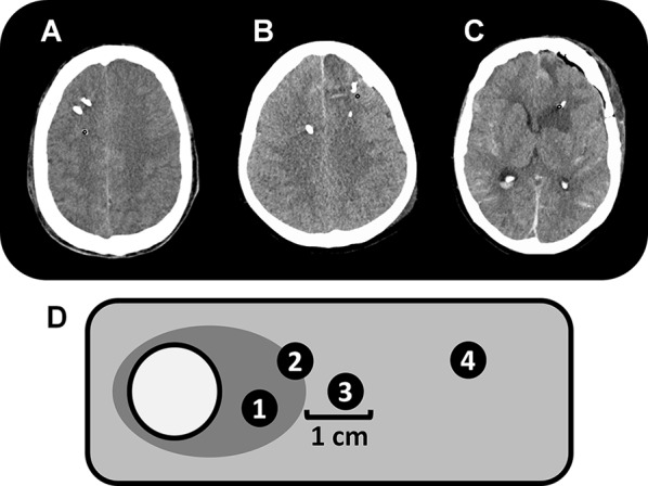
Fig. 1.

Examples for probe location in normal-appearing brain tissue (a), perilesional (b), and intralesional (c) probe location on axial computed tomography scans of the brain; panel d is a schematic diagram describing probe location classification. The white circle represents a parenchymal hemorrhage with perifocal edema (dark gray area), surrounded by normal-appearing brain tissue (light gray area). Tips of microdialysis probes completely surrounded (location 1) or in direct contact with (location 2) a hypodense area were classified as intralesional. Perilesional location was defined as lesion within 1 cm of the catheter tip (location 3). Location 4 represents normal-appearing brain tissue. The metabolic profile of locations 1, 2 and 3 were not different and summarized for comparison with location 4
