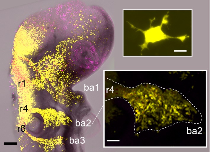
Fig. 1.

Neural crest cell migratory streams in the chick head co-labelled with pMES (EGFP) in yellow and DiI in purple. Rhombomeric segments of the hindbrain, from which some of the cranial NC cells emerge are labelled rhombomere 1 (r1), r4, and r6 with branchial arch target sites, branchial arch 1 (ba1), ba2, ba3 and the scalebar is 200 microns (black). The top inset is of a typical individual migrating neural crest cell with scalebar of 10 microns (white). The bottom inset is the neural crest cell migratory stream adjacent to r4 and the scalebar is 100 microns (white) (color figure online)
