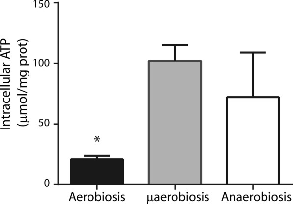
Fig. 3.

Intracellular ATP concentrations in S. epidermidis grown at different [O2]. Cells were grown at different [O2] in LB plus glucose. Cytoplasmic extracts were obtained from each of these cultures and used to measure intracellular ATP. ATP concentration was estimated using luciferase and interpolating into a standard curve (see “Materials and methods”). The average of three experiments is shown with SD. * indicates significant difference P < 0.05
