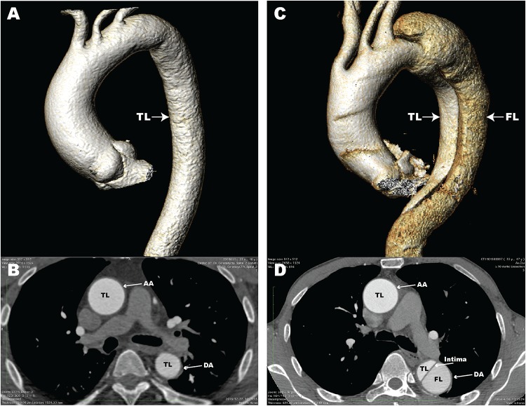
FIGURE 1.
Computed tomography angiography (CTA) images of a normal aorta and an aortic dissection (AD). (A) 3D reconstructed CTA image of a normal aorta; (B) Axial CTA image of a normal aorta with blood flow only in the true lumen (TL); (C) 3D reconstructed CTA image from a patient with AD; (D) Axial CTA image of AD with blood flow in both the true lumen and false lumen (FL). The aortic intima is located between the TL and FL. AA, ascending aorta; DA, descending aorta.

