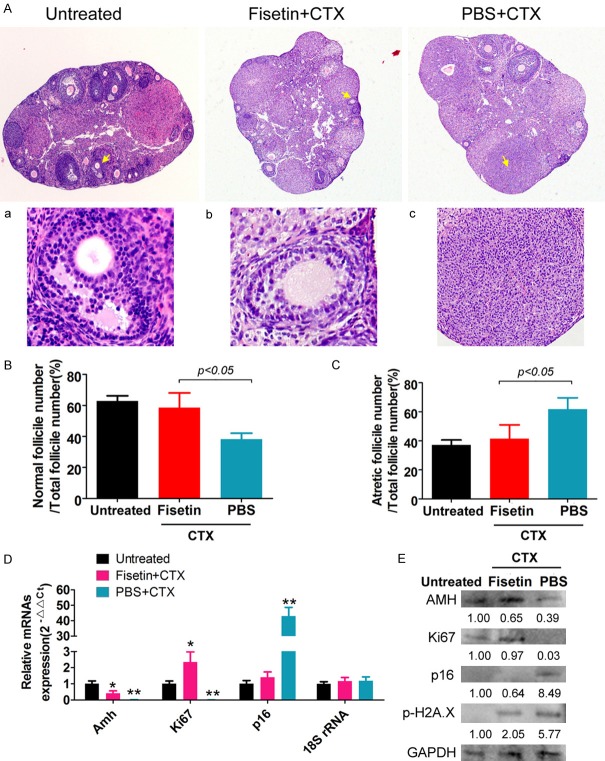
Figure 1.
Fisetin effectively alleviates ovarian damage in POF mice. (A) HE staining results of the ovarian tissues in various groups of mice (magnification: 100×). (a-c) are the high magnification images (400×) of the sites indicated with yellow arrows. (B) The proportion of normal ovarian follicles in the different groups of mice. (C) The proportion of atretic ovarian follicles in the different groups of mice. Pathological testing by HE staining showed that the number of atretic ovarian follicles significantly increased, while the number of quality ovarian granulosa cells significantly decreased in the ovarian tissues of POF mice. Ovarian follicles at various stages of development appeared in the ovaries of POF mice in the fisetin group. (D) qPCR for quantitation of mRNA levels of genes related to ovarian granulosa cell proliferation and aging. **P<0.01 vs PBS+CTX group, *P<0.05 vs PBS+CTX group, as calculated by t-test. (E) Western blot analysis to detect the expression levels of proteins related to ovarian granulosa cell proliferation and aging. qPCR and western blotting results revealed that the expression levels of p16 and pho-H2A.X in the ovaries of POF mice in the fisetin group were decreased significantly, while those of AMH and Ki67 were increased significantly.

