Figure 1.

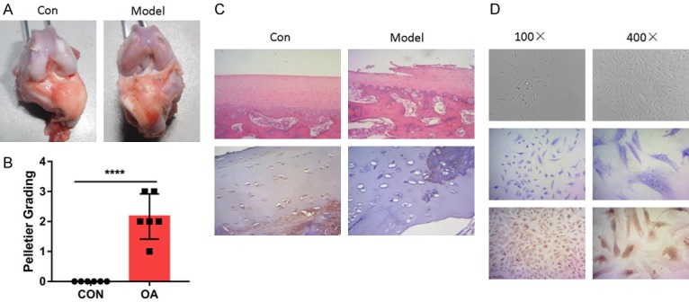
Rabbit OA model was successfully established for chondrocytes isolation. A. Macroscopic observations of rabbit articular cartilage in indicated group. Typical changes of the cartilage lesions were illustrated. B. Modeling was evaluated using Pelletier grading. C. Hematoxylin-eosin (HE) staining and immunohistochemistry (IHC) staining verified the proper modeling of OA in rabbit. The IHC-staining was conducted with the anti-Collagen II antibody. D. Isolated chondrocytes was evaluated using Microscopic observation, HE and IHC staining.
