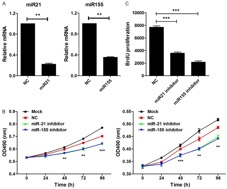
Figure 2.

Knockdown of miR-21 and miR-155 suppress cell proliferation. A. Raji cells were transfected with miR-21 inhibitor, miR-155 inhibitor, or negative control (NC) for 48 hours. The expression of miR-21 and miR-155 were measured by qPCR, wh normalized to human actin (n=3). B. Raji cells were transfected with miR-21 inhibitor, miR-155 inhibitor or negative control (NC) for 24 hours. Then, BrdU was added and incubated for another 24 hours. Cell proliferation was analyzed by flow cytometry. C. Raji cells were transfected with miR-21 inhibitor, miR-155 inhibitor, or negative control (NC) for 48 hours. Then, the CCK-8 assay (right) was performed to examine cell proliferation at various time points (0 h, 24 h, 48 h, 72 h, 96 h). Results are shown as absorbance at 490 nm. Data are representative of three independent experiments and shown as the mean ± SD. (**P<0.01, ***P<0.001, Student’s t-test).
