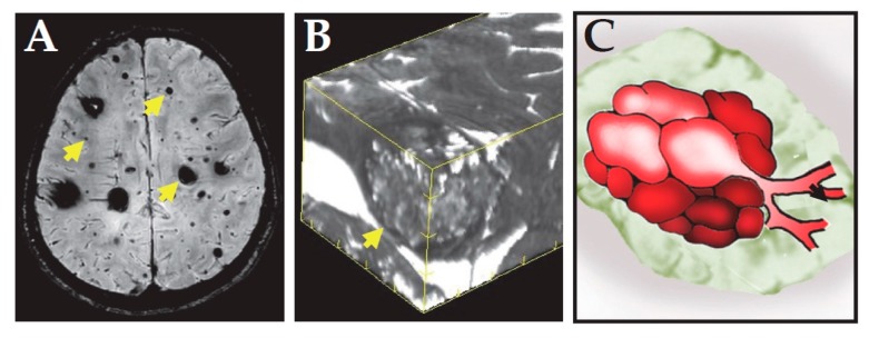
Figure 1.
Radiological presentation of CCM. (A) MRI image of the brain of a familial CCM patient. Susceptibility weighted imaging showed multiple dark CCM lesions with various sizes. Arrows indicate representative lesions. (B) 3D reconstruction of T2 weighted imaging of a CCM lesion. It shows the lesion is not uniform, but with popcorn appearance. The arrow indicates the location of the lesion. (C) Schematic presentation of a CCM lesion showing it is composed of nested dilated microvessels.

