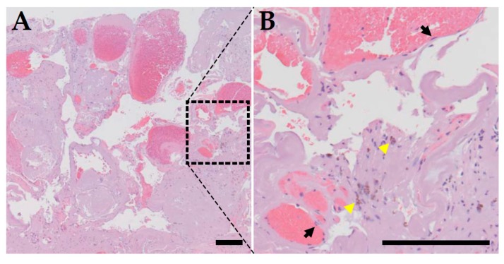
Figure 2.
Histopathological presentation of CCM. (A) H&E staining of a surgically resected CCM lesion. It is composed of clusters of thin walled dilated microvessels with no supporting smooth muscle cells beneath the endothelial cell layer and no intervening brain parenchyma. Thrombi are present within the lumen of capillaries within the CCM lesion. (B) High power image of the boxed region of panel A. Black arrows point to individual endothelial cells lining the inner surface of dilated capillaries, and yellow arrowheads point to hemosiderin deposition adjacent to the capillaries, a sign of chronic bleeding. Bar = 200 μm.

