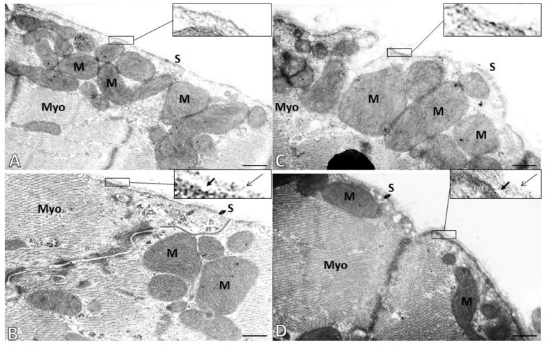
Figure 4.
Electron microscopic images demonstrate cardiomyocyte cell membrane (sarcolemma, S) integrity in the male (left panel A,B) and female (right panel, C,D) hypertensive rats without (A,C) or with omega-3 intake (B,D). Notice the pronounced degradation of the basement membrane of sarcolemma in non-treated male and female rat hearts (A,C). While preservation of basement membrane containing lamina lucida (short arrows) and lamina densa (long arrows) is seen in omega-3-treated rats (B,D). M—Mitochondria; Myo—Myofibrils. Scale bar—0.5 µm.

