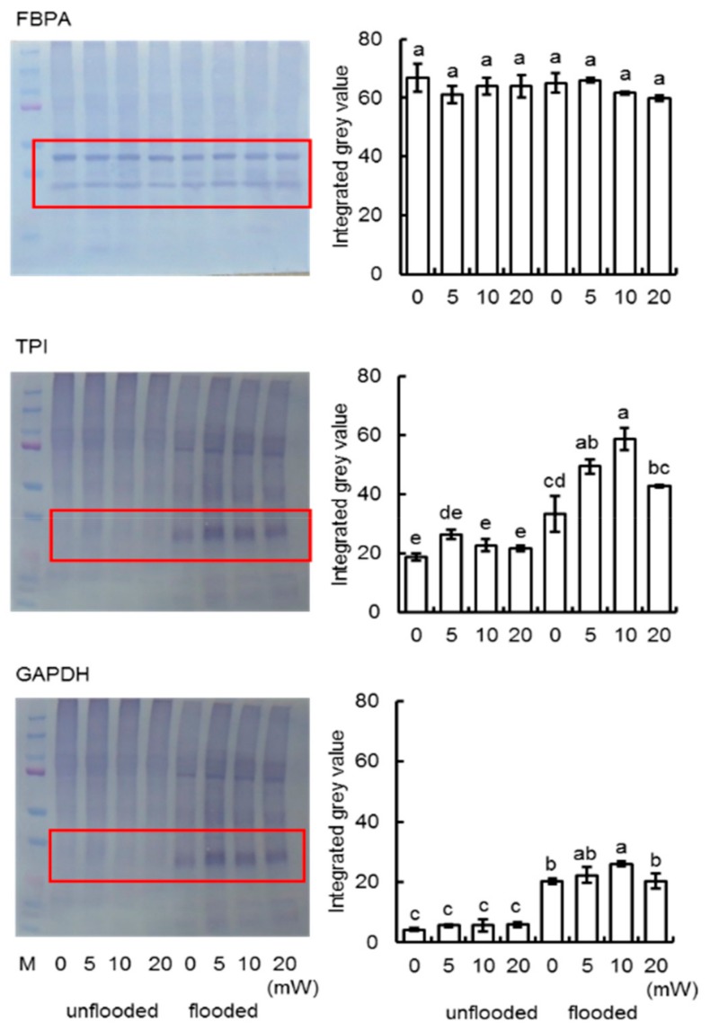
Figure 8.
Immunoblot analysis of proteins involved in glycolysis pathway. Proteins were extracted and separated on 10% SDS-polyacrylamide gel by electrophoresis and transferred onto membranes. The membranes were cross-reacted with anti-FBPA, anti-TPI, and anti-GAPDH antibodies. CBB staining pattern were used as loading control (Figure S5). The integrated densities of bands were calculated using ImageJ software. Pictures shows three independent biological replicates (Figure S7). Data are shown as means ± SD from three independent biological replicates.

