Figure 4.
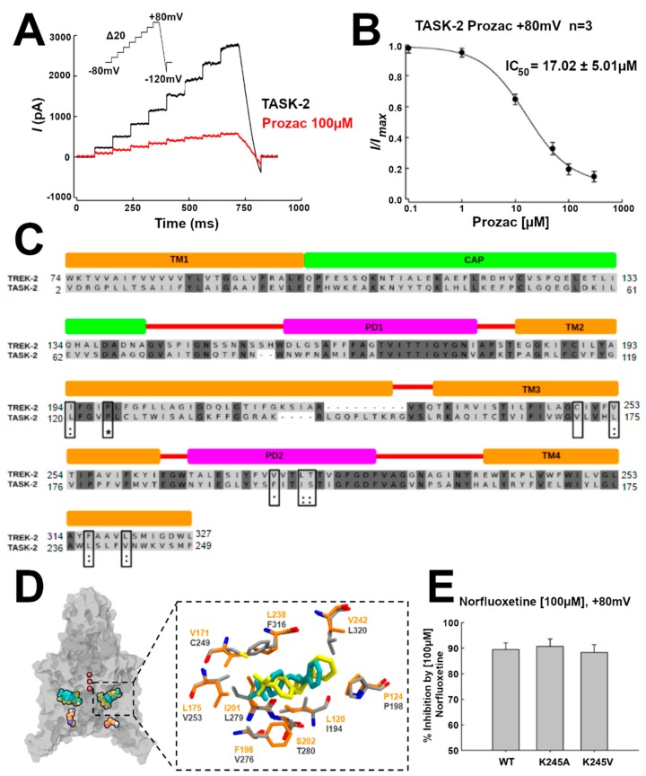
TASK-2 is blocked by Prozac. (A) Whole-cell recordings showing the effect of Prozac (100 µM) on TASK-2 channels. The currents were evoked using the voltage protocol from −80 to +80 mV (see inset); (B) Concentration–response curve with the IC50 value of TASK-2 inhibition by Prozac. The block was analyzed at the end of the test pulse at +80 mV; (C) Docking of Prozac in the T2Tre2OO structure (yellow) shows a similar position as Prozac in the TREK-2 crystal structure (cyan). Prozac binding site residues in TREK-2 are shown in gray, whereas the homologous residues in TASK-2 are in orange. The protein is shown in transparent surface representation. The ions in the selectivity filter are red. K245 residues are shown as reference in orange; (D) Pairwise sequence alignment between TREK-2 and TASK-2 channel [27]. Prozac binding site in TREK-2 is composed by residues of TM2, TM3, PD2, and TM4. They are conserved in TASK-2 channel. * Identical residues; : very conserved residues; ∙ conserved residues; (E) Percentage of inhibition by 100 µM Prozac of wild-type TASK-2 and two mutants of K245 residue. Results are shown as means ± Standard Error of the Mean (SEM).

