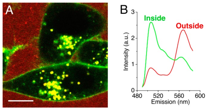
Figure 6.
(A) Confocal fluorescence image of KB cells incubated with DiO/DiI loaded micelles. The image shows the loss of Förster resonance energy transfer on the cell surface and intracellular space. (B) Normalized spectra of the measured fluorescent signal outside (red) and inside (green) the cells. (Scale bar: 10 µm.) (Adapted from Reference [49], Copyright 2008 National Academy of Sciences).

