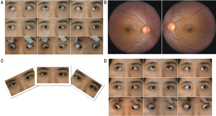
Figure 1. Clinical photograph of case 1.
A: A 9-gaze eye position photograph in a 26-year-old female with congenital left IOP presenting with underelevation in adduction, 20 PD esotropia and 15 PD hypotropia of the left eye in the primary position. The difference of esotropia in upward and downward gaze of 25° was 14 PD (A pattern esotropia). B: Fundus photograph showing incyclotorsion in her right eye. C: Although head position appeared normal, with a head tilt test she demonstrated an increased vertical deviation when her head was tilted to the right side. D: Orthophoria was present in the primary position and her A pattern esotropia dissipated at 12mo after tenotomy of the superior oblique and a 3 mm medial rectus recession in the left eye.

