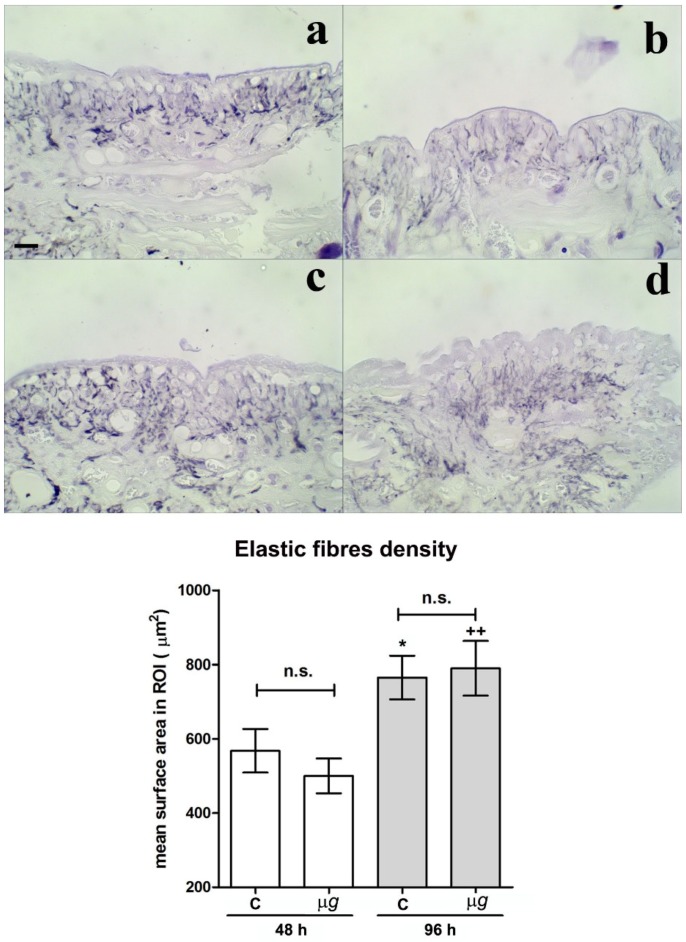
Figure 3.
Effect of modeled µg on in vivo model of sutured wound healing (Hirudo medicinalis): elastic fibre content in the connective tissue at the wound site. Light microscopical features and morphometric quantitation of paraldehyde fuchsin-stained elastic fibres at the wound site after 48 h and 96 h exposure to modeled µg (b,d), compared with 1× g controls (a,c). In the graph C = 1× g; bar = 20 µm; * p < 0.05 vs. C-48h, ++ p < 0.01 vs. µg-48 h, (n = 3); n.s. = not significant.

