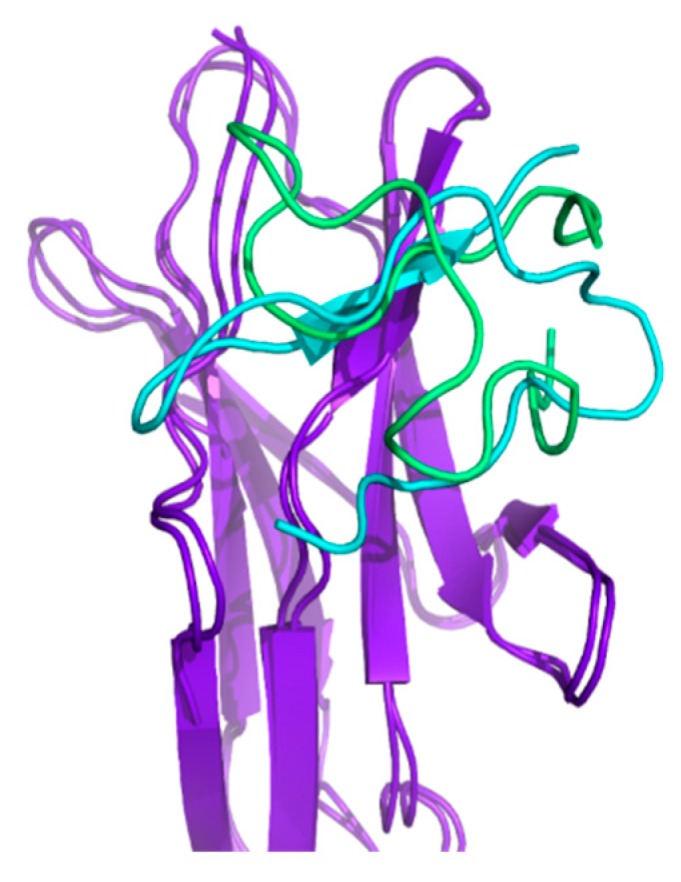
Figure 4.

The comparison between the structure of the dominant cluster of HVEM (14–39) peptide docking to BTLA (purple) obtained from (i) simulations with peptide restraints based on the crystal structure (cyan) of the BTLA/HVEM complex and (ii) simulations with peptide restraints based on the structure derived from the nuclear magnetic resonance (NMR) spectra (green).
