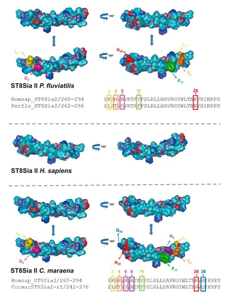
Figure 8.
Three-dimensional (3D) structure of PSTD motifs in fish ST8Sia II. The 3D model of human ST8Sia II PSTD in addition to PSTD from P. fluviatilis and C. maraena was simulated, based on the 3D model of human ST8Sia IV PSTD (Protein Data Bank entry 6AHZ) using YASARA. The electrostatic potential surfaces are displayed. The exchanged amino acids are colored in an additional version of the 3D structure to highlight the position of the exchange: K2 → L2 (yellow), H4 → T4 (orange), N6 → D6 (magenta), Y11 → F11 (green), and K28 → N28 (red) for P. fluviatilis and K2 → L2 (yellow), H4 → T4 (orange), N6 → D6 (magenta), R8 → H8 (violet), Y11 → F11 (green), K28 → N28 (red), and H30 → Q30 (light blue) for C. maraena. For the determination of the 3D structure of human ST8Sia IV PSTD, a peptide with one additional amino acid on the N-terminus and two additional amino acids on the C-terminus of PSTD were used [66].

