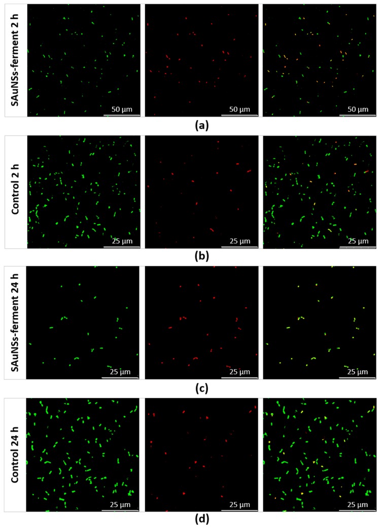
Figure 7.
Confocal laser scanning microscopy images of SAuNPs–ferment after 2 h (a) and 24 h (c) incubations compared to control cultures lacking gold ions at the same times: 2 h (b) and 24 h (d). From left to right for each sample: all bacteria (spots stained green by SYTO9), dead bacteria (spots stained red by propidium iodide (PI)) and a merge of both images, where green and yellow spots corresponds to live and dead bacteria, respectively.

