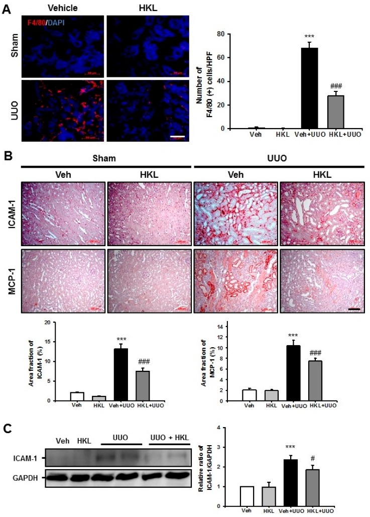
Figure 3.
Effect of honokiol on unilateral ureteral obstruction (UUO)-induced renal inflammation. (A) Representative sections of kidneys from sham- and UUO-operated mice treated with vehicle (Veh) or honokiol (HKL) were immunofluorescence-stained with F4/80 (red). The nucleus was stained with DAPI (blue). Scale bar = 50 μm. The bar graph shows the number of F4/80-positive cells (%) in the sham and UUO kidneys from ten randomly chosen, non-overlapping fields at a magnification of 400× (n = 10 per group). (B) Representative sections of kidneys from sham- and UUO-operated mice treated with Veh or HKL were stained with ICAM-1 and MCP-1. Scale bar = 100 μm. The bar graph shows area fractions (%) of ICAM-1 and MCP-1 in the sham- and UUO-operated kidneys from ten randomly chosen, non-overlapping fields at a magnification of 400× (n = 10 per group). (C) Representative Western blot analysis of ICAM-1 protein expression in the kidneys from sham- and UUO-operated mice treated with Veh or HKL. The bar graph shows the densitometric quantification presented as the relative ratio of each protein to GAPDH. The relative ratio measured in the kidneys from sham-operated mice treated with Veh is arbitrarily presented as 1. Data are expressed as mean ± SD. *** p < 0.001 versus Veh or HKL; # p < 0.05 and ### p < 0.001 versus UUO treatment with Veh. Sham, sham-operated mice; UUO, unilateral ureteral obstruction; Veh, vehicle; HKL, honokiol; ICAM-1, intercellular adhesion molecule-1; MCP-1, monocyte chemoattractant protein-1; DAPI, 4’,6-diamidino-2-phenylindole; GAPDH, glyceraldehyde 3-phosphate dehydrogenase.

