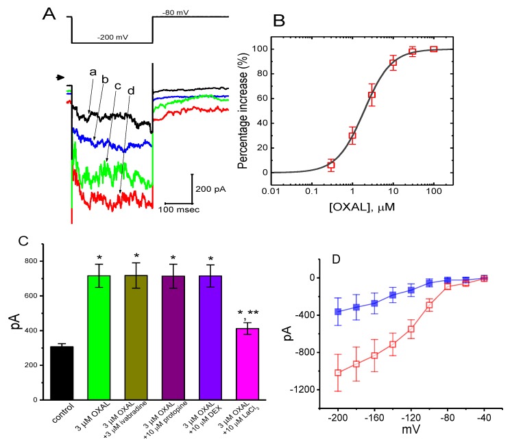
Figure 5.
The stimulatory effect of OXAL on membrane electroporation-induced inward current (IMEP) in GH3 cells. Cells were immersed in Ca2+-free Tyrode’s solution and the examined cells were maintained at −80 mV. (A) Representative IMEP traces elicited by membrane hyperpolarization from −80 to −200 mV (as indicated in the upper part). Arrowhead is zero current level; a: control; b: 1 μM OXAL; c: 3 μM OXAL; d: 10 μM OXAL. (B) Concentration-dependent stimulation of IMEP by OXAL (mean ± SEM; n = 8 for each point). (C) Summary bar graph showing effects of OXAL, OXAL plus ivabradine, OXAL plus protopine, OXAL plus DEX (dexmedetomidine), and OXAL plus LaCl3 (mean ± SEM; n = 9 for each bar). Current amplitude was taken at the end of hyperpolarizing pulse from −80 to −200 mV. The smooth line represents least-squares fit to a Hill function detailed in Materials and Methods. * Significantly different from control (p < 0.05) and ** significantly different from 3 μM OXAL alone group (p < 0.05). (D) Averaged I–V relationships of IMEP obtained in the absence (closed squares) and presence (open squares) of 3 μM OXAL (mean ± SEM; n = 9 for each point). As the whole-cell mode was firmly established, the cells were maintained at −80 mV and a family of voltage pulses ranging between −200 and −40 mV with a duration of 300 ms at increments of +20-mV were applied. Current amplitude was measured at the end of each voltage pulse.

