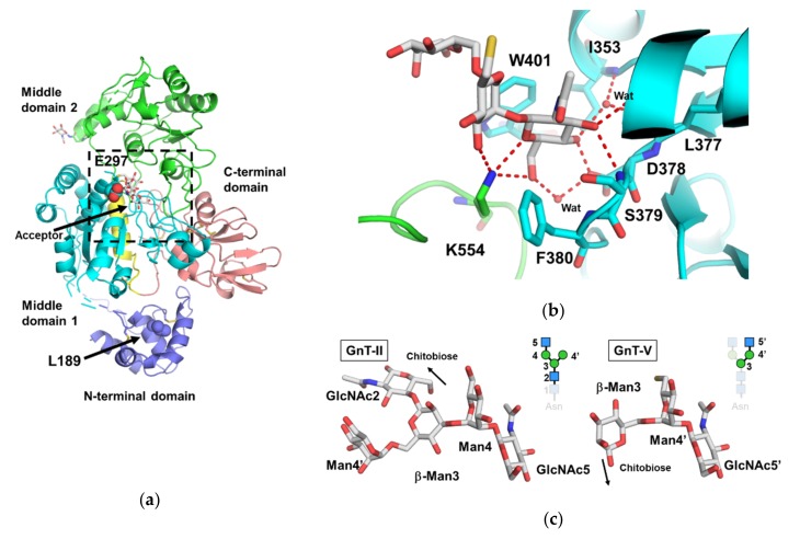
Figure 4.
Crystal structure of human GnT-V. (a) Overall structure of the human GnT-V luminal domain (PDB code: 5ZIB). The N-terminal domain, middle domain 1, 2, and C-terminal domain are colored in blue, cyan, green, and pink, respectively. The short insertion in middle domain 1 is colored in yellow. The position of the acceptor N-glycan was obtained by superposition of the mini-GnT-V-glycan complex (PDB code: 5ZIC). Two important residues (L189 and E289) denoted in the text are shown as sphere models. The oxygen atoms of E289 are highlighted in red. The putative catalytic center is highlighted in the black dotted box. (b) Close up view of the acceptor N-glycan binding site (PDB code: 5ZIC). The direct and water-mediated hydrogen bonds are shown in red dotted lines. Two aromatic residues (F380 and W401), which may determine the branch specificity of GnT-V, are also shown. (c) Structural comparison between the GnT-II acceptor complex and GnT-V acceptor complex. The superposition is based on GlcNAc residues of both branches (GnT-II: α1-3 branch, GnT-V: α1-6 branch). The disaccharide units (GlcNAcβ1-2Man) of the two structures are well superimposable.

