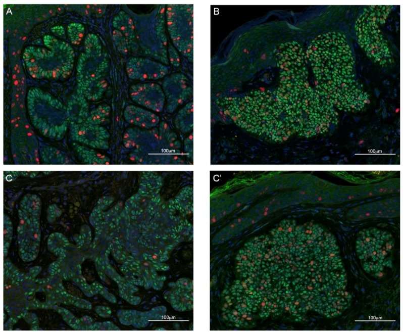
Figure 2.
Examples of immunofluorescence staining of sections from skin biopsies of BCC with indolent growth. TBX1 in green, Ki67 (a cell proliferation marker) in red, and DAPI (DNA stain) in blue. (A) Example of polarized distribution of TBX1+ cells, defined as localization of positive cells at the periphery of the lesion (palisade cells), while internal cells are mostly TBX1-negative; (B) example of diffuse distribution, defined as localization of positive cells to the entire lesion. (C,C′) represent different regions of the same sample; note that both types of distribution, polarized (C), and diffuse (C′) are present. Scale bar 100 µm. See Figure S1 for examples of hematoxylin-eosin staining of these biopsies.

