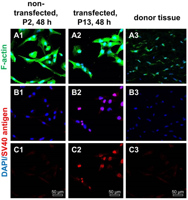
Figure 2.
F-actin and SV40 T antigen expression in non-transfected and transfected human ACL ligamentocytes after 48 h in 2D culture as well as native tissue. (A1–C1) Non-transfected ligamentocytes, passage (P) 2, (A2–C2) ligamentocytes transfected with the SV40 plasmid (P13) and the original donor tissue in situ (A3–C3). (A1–A3) Cells were stained with phalloidin-Alexa488 (green) to depict the F-actin cytoskeleton. (B1–B3) Cells were immunolabeled with a specific anti-SV40 antibody (red), the cell nuclei were counterstained with 4′,6′-diamidino-2-phenylindol (DAPI, blue) (A1–B3). Scale bars: 50 µm.

