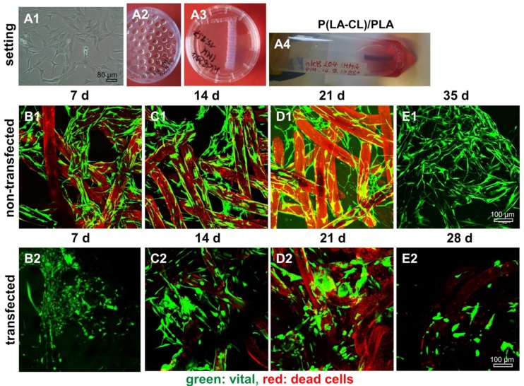
Figure 8.
Representative microscopic fields of non-transfected and SV40 transfected ACL ligamentocytes on P(LA-CL)/PLA scaffolds. Experimental design, cell expansion (A1); self-assembly using hanging drop method (50,000 cells per spheroid) (A2); macroscopic image of wet P(LA-CL)/PLA scaffolds (A3); and dynamical rotatory scaffold culture (A4). (B1–E2) Live/Dead staining of cultures maintained up to day 35 (non-transfected cells, (B1–E1)) and day 28 (SV40 transfected cells, (B2–D2)). Vital cells: Green. Dead cell: Red. Scaffold fibers: Slightly red colored. Three independent experiments were performed. Scale bars: 100 µm.

