Figure 1.
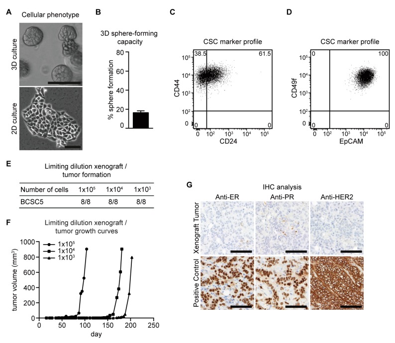
Characterization of BCSC5 in vitro and in vivo. (A) Representative pictures of BCSC5 cultured in 3D and 2D conditions, scale bar 100 μm. (B) Sphere-forming capacity of BCSC5 cells in an anchorage-independent growth assay (n = 3). Data represent means + SEM. (C,D) Expression patterns of CD24 and CD44 (C) as well as EpCAM and CD49f (D) in BCSC5 cells analyzed by flow cytometry. (E) Tumor formation in limiting dilution xenografts of BCSC5. (F) Representative growth curves for limiting dilution assay of BCSC5 xenografts in immunocompromised NOD/SCID mice. (G) Immunohistochemical (IHC) analysis of ER, PR, and HER2 on sections of the BCSC5 xenograft tumors, scale bar 100 μm.

