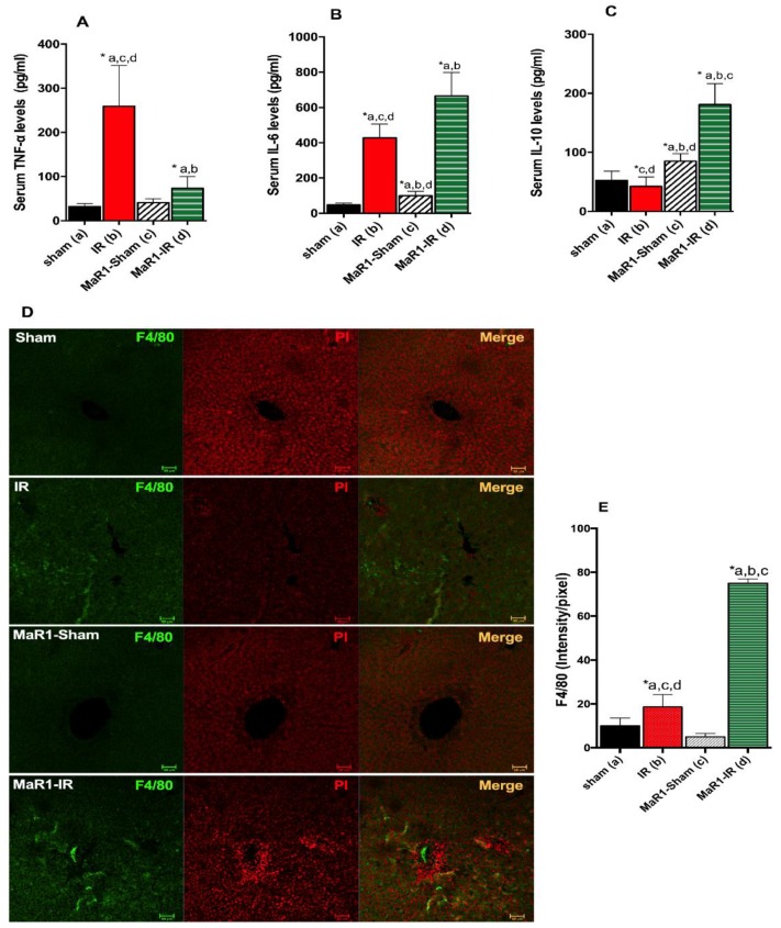
Figure 6.
Effect of MaR1 on inflammatory response. Serum levels of inflammatory cytokines (A) tumor necrosis factor (TNF)-α, (B) interleukin (IL)-6 and anti-inflammatory cytokine, and (C) IL-10 levels after IR were quantified. (D) Representative images depicting hepatic macrophages stained with the F4/80 marker in green and nuclei in red (propidium iodine), assessed by immunofluorescence in paraffined fixed liver tissues. (E) The fluorescence intensity was evaluated. The plots are represented as mean ± SEM, n = 4 rats per experiment, Image fields measured per rats >6. Significance was assessed by one-way ANOVA and the Tukey’s post-test. Asterisk indicates p < 0.05, and the letters identify the experiments that are compared and present this statistical difference. Scale bar indicates 50 µm.

