Figure 5.
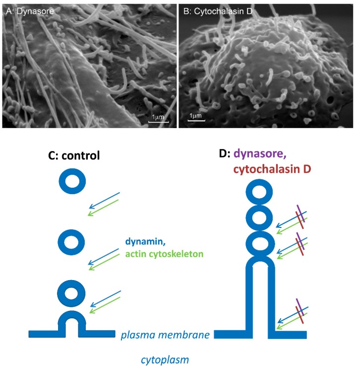
Scheme of the cytoneme formation. (A,B) Scanning electron microscopy images of the surface of neutrophils that were attached to fibronectin-coated substrata during 20 min in the presence of 200 µm dynasore (A) or 10 µm cytochalasin D (B). (C,D) GTPase dynamin, in collaboration with an intact actin cytoskeleton separates exocytotic vesicles from the plasma membrane (C). Inhibition of dynamin with dynasore and/or depolymerization of actin filaments with cytochalasin D block the cleavage of exocytotic vesicle from the plasma membrane and from each other, and the secretory process extends from the cells as tubulovesicular cytonemes (D). The photographs shown in this figure are similar to the photographs published in our previous article [9].

