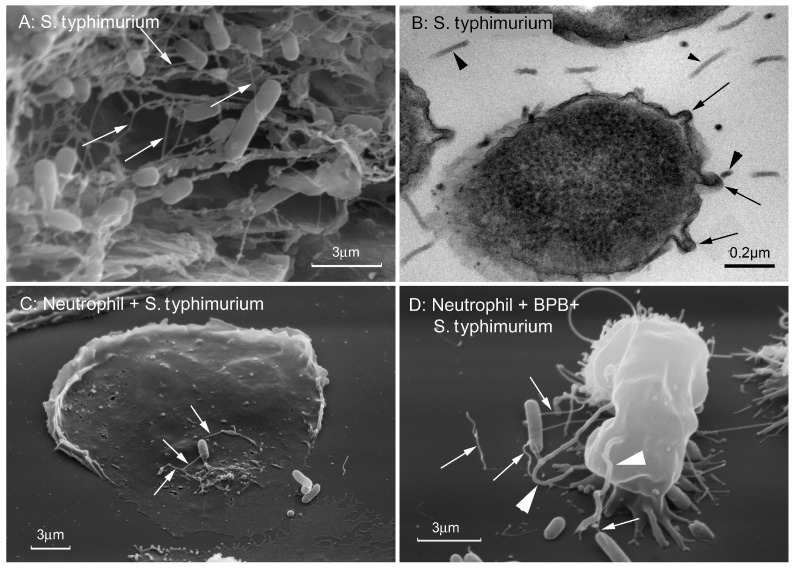
Figure 6.
Cytonemes of Salmonella typhimurium. (A) Scanning electron microscopy images of S. typhimurium biofilm grown on gallstones. White arrows indicate 60 nm wide cytonemes interconnecting bacteria in biofilm. (B) Transmission electron microscopy images of a thin section of S. typhimurium biofilm grown on agar. Black arrows indicate fragments of 60 nm-wide outer membrane tubular extensions (cytonemes) of bacteria. Black arrowheads indicate 25 nm in diameter bacterial flagella. (C,D) Scanning electron microscopy images of S. typhimurium attached to the control (C) and 4-bromophenacyl bromide (BPB)-treated (D) neutrophils. White arrows indicate the 60 nm-wide bacterial cytonemes. White arrowheads indicate the cytonemes of neutrophils with a diameter of 200 nm. The photographs shown in this figure are similar to the photographs published in our previous article [18].

