Figure 1.
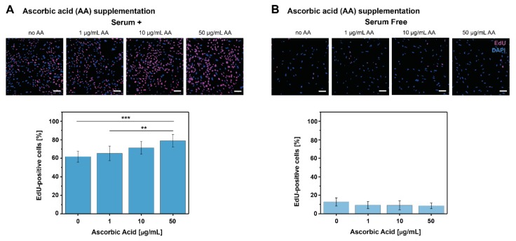
Effect of AA on tenocyte proliferation. Representative CLSM images from the EdU proliferation assay and EdU-positive cells [%] for each AA concentration (μg/mL) tested in (A) serum+ conditions and (B) serum-free conditions. The effect of different PDGF-BB concentrations on tenocyte proliferation assessed by the EdU assay is presented in Figure S1. The total EdU-positive cells [%] was calculated as EdU positive cells (pink) in relation to the total cell number (DAPI stained, blue). Scale bars: 100 μm. (Data is shown as mean ± standard deviation, ** p < 0.01, *** p < 0.001 obtained by one-way ANOVA).

