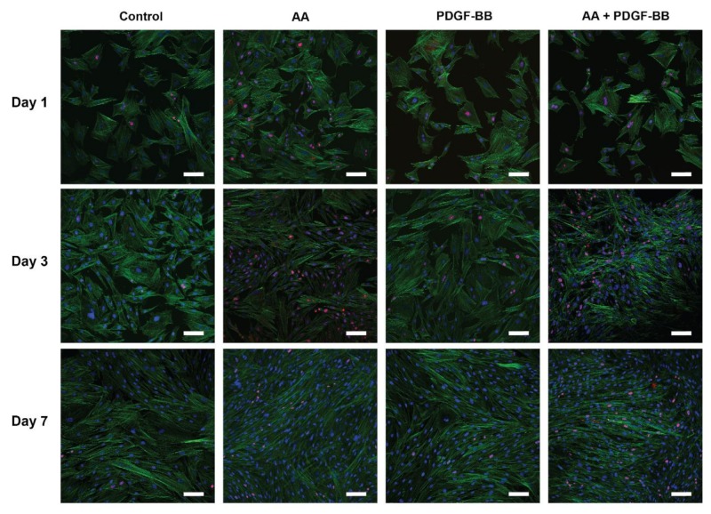
Figure 2.
Effect of AA and PDGF-BB on tenocyte proliferation and morphology in cell culture. Representative CLSM images of tenocytes cultured in serum+ medium for a period of 7 days and supplemented with either AA (10 μg/mL), PDGF-BB (25 ng/mL), combination of AA (10 μg/mL) + PDGF-BB (25 ng/mL), or untreated (control). Cells were stained with phalloidin (green) for visualizing the cytoskeleton, ki-67 (red) as a proliferation marker, and DAPI (blue). Scale bars: 100 μm.

