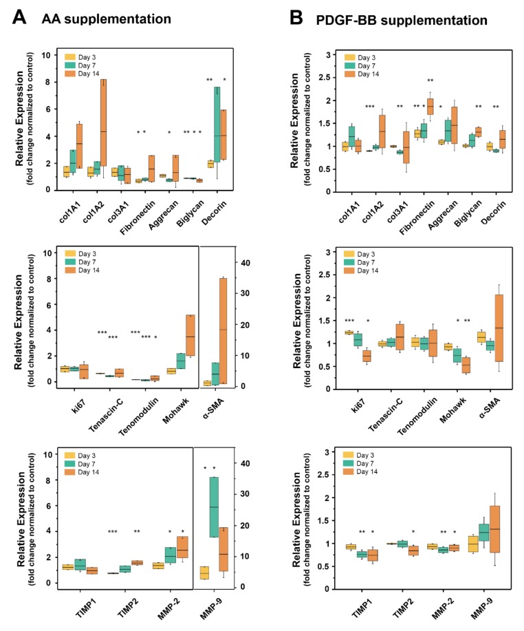
Figure 3.
Gene expression of tenocytes supplemented with AA or PDGF-BB over 14 days in 2D cell culture. (A) Box-plots of time-dependent (day 3, 7 and 14) relative gene expression in tenocytes supplemented with AA (50 μg/mL), normalized to non-stimulated tenocytes and (B) box-plots of time-dependent (day 3, 7 and 14) relative gene expression in tenocytes supplemented with PDGF-BB (25 ng/mL), normalized to non-stimulated tenocytes. In the experiments with AA, tenocytes isolated from three different rabbits were used (biological replicates n = 3), while for PDGF-BB experiments, cells from four different rabbits were used (biological replicates n = 4). Technical replicates in cell culture were performed in triplicate and in PCR experiments in duplicate. Significant differences were based on unpaired t-tests for each time point (whiskers on the box-plots represent standard deviation, * p < 0.05, ** p < 0.01, *** p < 0.001).

