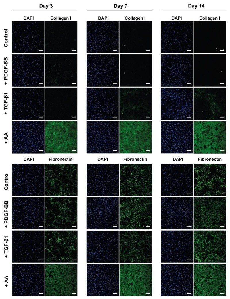
Figure 4.
Extracellular matrix deposition by tenocytes stimulated with PDGF-BB, AA or TGF-β1. Representative CLSM images of collagen I or fibronectin staining (green) and cell nuclei DAPI staining (blue) of rabbit tenocytes cultured in vitro in 2D, treated with PDGF-BB (50 ng/mL), TGF-β1 (10 ng/mL), AA (50 μg/mL) or untreated (control) on day 3, 7 and 14. For additional data of the same experiment performed with tenocytes isolated from two other animals and for quantification of the fibronectin deposition under the different treatments, we refer to the figures in the supporting information. Scale bars: 100 μm.

