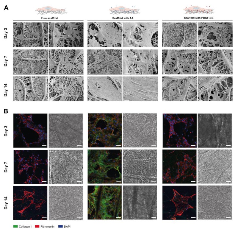
Figure 5.
Tenocytes cultured on pure and bioactive scaffolds over a period of 14 days. (A) SEM images of tenocytes, seeded on bioactive electrospun DP scaffolds, containing either AA, PDGF-BB (bioactive scaffolds) or none of them (pure scaffold). The cell morphology and ECM deposition overtime is observed on the SEM images. (B) Representative CLSM images of collagen I (green), fibronectin staining (red) and cell nuclei DAPI staining (blue) of rabbit tenocytes cultured onto the electrospun scaffolds. Scale bars: (A) 20 µm and (B) 50 μm.

