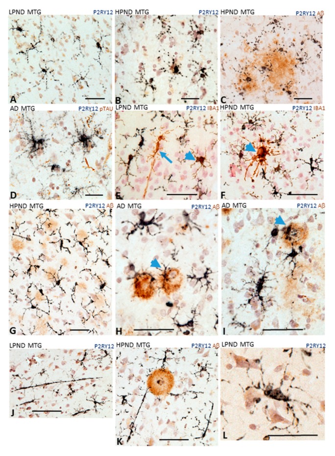
Figure 5.
Different microglial morphologies associated with P2RY12 expression. Representative immunohistochemistry results of tissue sections stained to identify P2RY12 (purple) alone and Aβ (brown), IBA-1 or phosphorylated tau. (A). Ramified microglia in LPND case. (B,C). Microglia with fragmented morphology in HPND cases. (C). Fragmented microglia associated with diffuse Aβ plaques. (D). P2RY12 microglia with tufted morphology in AD case. (E,F). Colocalization of P2RY12 and IBA-1. (E). Rod shaped IBA-1-positive microglia (brown arrow) with minimal P2RY12 immunoreactivity. P2RY12-positive, IBA-1-positive microglia (arrowheads (E,F)). All P2RY12 immunoreactive microglia showed some IBA-1 immunoreactivity. (F). IBA-1 positive cluster surrounded by P2RY12 microglia. (G–I). Different morphologies of P2RY12-positive microglia interacting with Aβ plaques. (G). P2RY12-positive microglia with long processes interacting with diffuse plaques in HPND case. (H,I). P2RY12-positve microglia with activated morphologies (large cell bodies, short processes) interacting with dense Aβ plaques. (J,K). P2RY12-positive rod-shaped microglia in LPND (J) and HPND (K) sections. (L) P2RY12-positive microglial processes show interactions with neurons. Scale bars represent 50 μm.

