Figure 1.
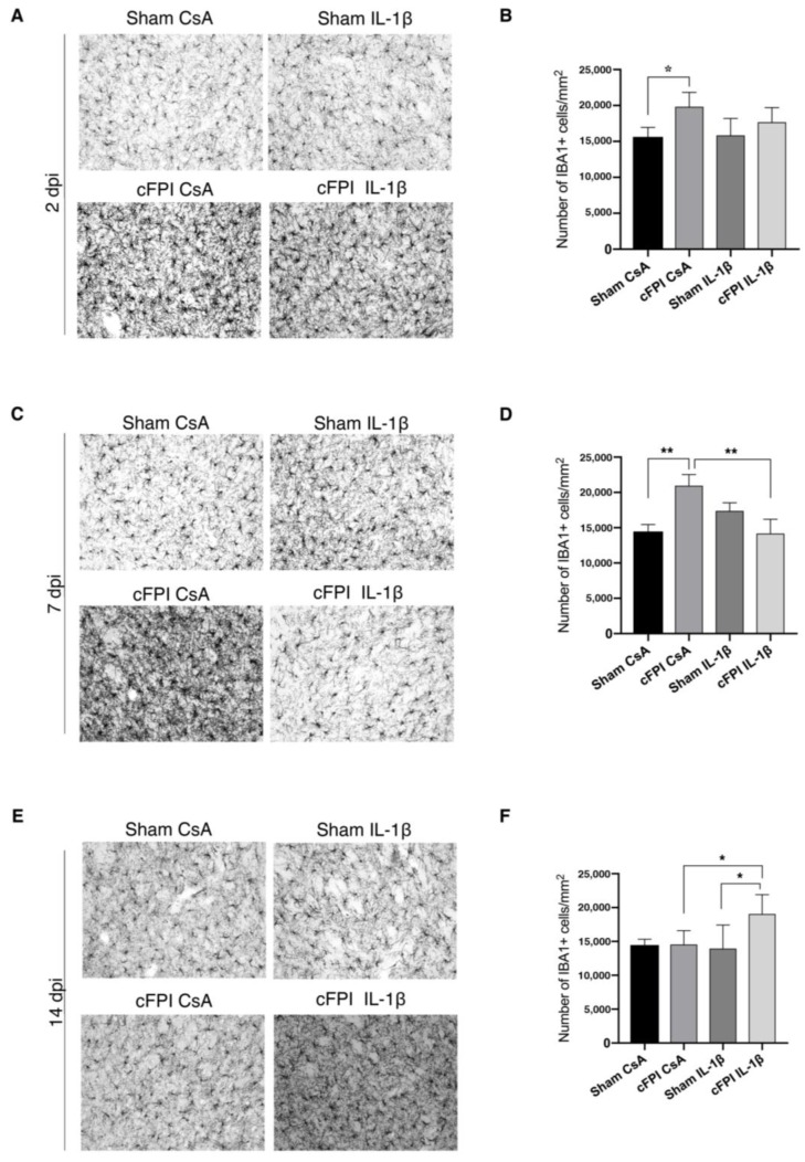
IL-1β neutralization attenuates the increase of Iba1+ microglia cells in the globus pallidus. Compared with sham CsA mice at two dpi, (A,B) the number of Iba1+ cells was increased in CsA cFPI animals (sham CsA, n = 4; sham IL-1β, n = 3; cFPI CsA, n = 6; cFPI IL-1β, n = 6; * p ≤ 0.05), while neutralizing IL-1 β had no effect on the number of Iba1+ microglial cells. At two dpi, Iba1 positive microglia processes were thin and ramified in the sham CsA group compared with thicker processes in the cFPI CsA animals (C,D) At seven dpi, the increase in the number of Iba1+ microglia cells persisted in the cFPI CsA animals compared with sham groups, which was significantly reduced by IL-1β neutralizing antibody treatment (sham CsA, n = 4 sham IL-1β, n = 3; cFPI CsA, n = 7; cFPI IL-1β, n = 6; ** p ≤ 0.01). Iba-1 positive microglia had thin and ramified processes in the cFPI IL-1β animals similar to sham CsA group (E,F) At 14 dpi, no difference was observed in the number of Iba1 positive microglia cells in the cFPI CsA animals in comparison to the sham groups (sham CsA and sham IL-1β). In the GP, the Iba-1 immunoreactivity was significantly increased by IL-1β neutralization in the injured mice (sham CsA, n = 3; sham IL-1β, n = 3; cFPI CsA, n = 8; cFPI IL-1β, n = 11; * p ≤ 0.05). Iba1 positive microglia cells had thicker processes in the cFPI IL-1β animals as compared to other groups. cFPI, central fluid percussion injury; dpi, days post-injury; CsA, inactive control antibody against cyclosporin A; IL-1β, interleukin 1 beta; Scale bars 20 μm.

