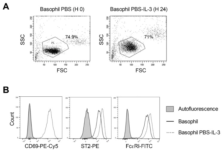
Figure 3.
Response of the phosphate buffered saline (PBS)-treated human peripheral blood basophils to IL-3 stimulation. Basophils were incubated on ice for 5 min with PBS. Cells were then washed and cultured in serum-free X-VIVO 15 medium (0.1 × 10⁶ cells/well per 200 µL) in 96-well U-bottomed plate along with IL-3 (100 ng/0.5 million cells) for 24 h. (A) The forward and side scatter plot of the basophils immediately following PBS treatment and following 24-h culture in the presence of IL-3. (B) Histogram overlays showing the expression of basophil activation markers CD69, ST2 and FcεRI in PBS-treated basophils cultured for 24 h in IL-3. Representative data from four donors are presented.

