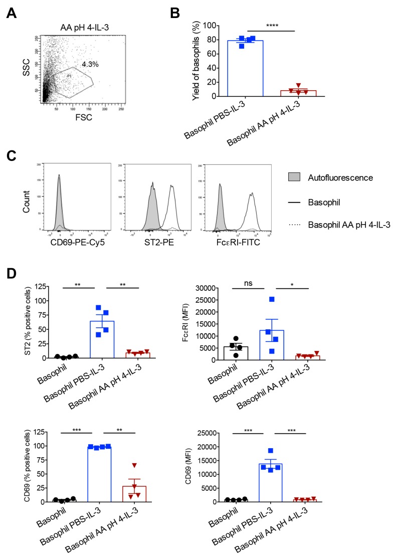
Figure 4.
Response of the acetic acid buffer (pH 4)-treated human basophils to IL-3 stimulation. Basophils were incubated on ice for 5 min either with phosphate buffered saline (PBS) or ice-cold acetic acid buffer pH 4 (AA pH 4). Cells were then washed and cultured in serum-free X-VIVO 15 medium (0.1 × 10⁶ cells/well per 200 µL) in 96-well U-bottomed plate along with IL-3 (100 ng/0.5 million cells) for 24 h. (A) The forward and side scatter plot of AA pH 4-treated basophils following 24-h culture in IL-3. (B) The yield of basophils (mean ± SD, n = 4 donors) after 24 h culture of basophils in IL-3. The values were calculated based on the percentage of cells in the P1 gate of forward and side scatter plot. (C) Histogram overlays showing the expression of basophil activation markers CD69, ST2 and FcεRI in AA pH 4-treated cells after 24 h culture in IL-3. Representative data from four donors are presented. (D) The expression (mean ± SD, n = 4 donors) of various activation markers (% positive cells or median fluorescence intensity, MFI) after 24 h culture of basophils in IL-3. * p < 0.05; ** p < 0.01; *** p < 0.001; **** p < 0.0001; ns, not significant; two-sided Students t-test (panel B) or one-way ANOVA with Tukey’s multiple comparison test (panel D).

