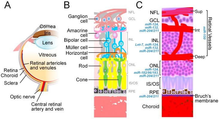
Figure 2.
The anatomy of the eye and relevant miRNAs. (A) The schematic diagram illustrates the main structures of the human eye. (B) The schematic representation of the cell types in the neural retina depicts their cellular connections (including ganglion cells, amacrine cells, bipolar cells, horizontal cells, as well as rod and cone photoreceptors) and supporting cells (Müller cells and RPE). (C) A cross section of the eye shows the laminar organization of the nuclear layers (GCL, INL, and ONL), the retinal vasculature, and segments of photoreceptors (IS/OS). The RPE monolayer, with Bruch’s membrane underneath, is located between the neural retina and the choroid complex. miRNAs that regulate the physiological functions or pathological conditions related to each retinal neuronal and vessel layers, and RPE, are listed next to respective histological structure. Deep, deep layer of retinal vessels; GCL, ganglion cell layer; INL, inner nuclear layer; Int, intermediate layer of retinal vessels; IS/OS, inner/outer segments; ONL, outer nuclear layer; RPE, retinal pigment epithelium; Sup, superficial layer of retinal vessels. Figure adapted from “Animal models of ocular angiogenesis: from development to pathologies” by Liu et al. 2017, FASEB J, 31(11), p. 4665–4681 [57].

