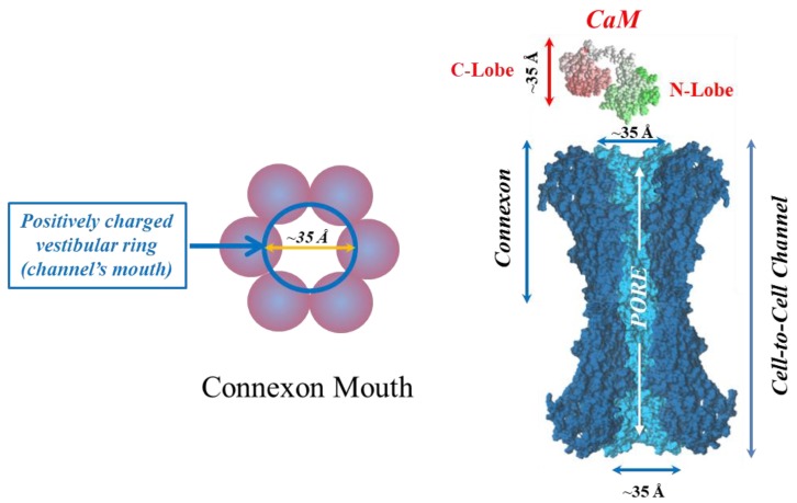
Figure 18.
Each negatively charged CaM lobe is ~25 × 35 Å in size (right panel), which is similar to the positively charged channel’s mouth. Thus, a CaM lobe could interact with the connexon’s mouth. In the right panel, the channel is split lengthwise so that the actual pore diameter (light blue area) is visible along the entire channel. CaM and connexons images (right panel) were generously provided by Drs. Francesco Zonta and Mario Bortolozzi (Venetian Institute of Molecular Medicine, VIMM, University of Padua, Italy).

