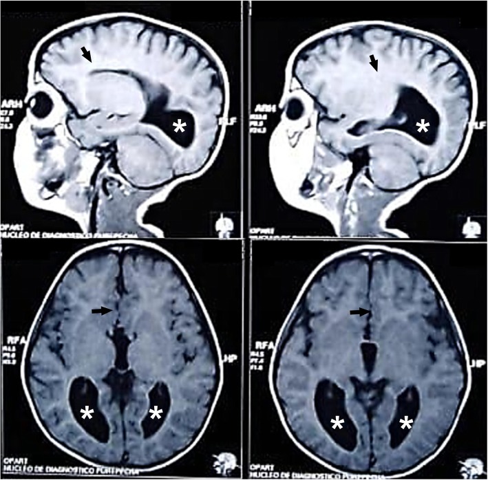
Fig. 1.
Brain magnetic resonance imaging images of the clinical case. Sagittal (upper panels) and transversal (lower panels) T1-weighted brain magnetic resonance imaging images showing total agenesis of the corpus callosum (black arrows) and enlargement of the occipital horns of the lateral ventricles (white stars).

