
Fig. 2.
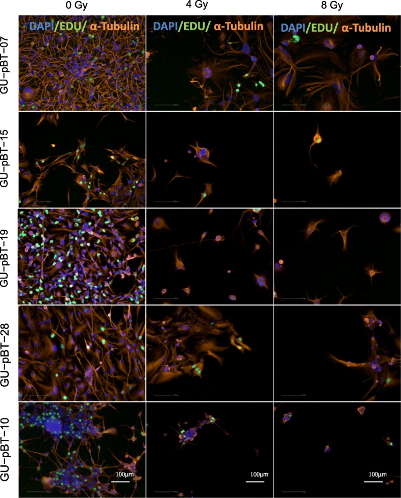
Radiation-induced cellular and nuclear morphological changes. Immunocytochemistry showing cellular morphology with α-tubulin-stained cytoskeleton (orange), DNA-synthesizing cells by EdU incorporation (green) and nuclear staining with DAPI (blue) at normal culture conditions (0 Gy) and exposed to radiation (4 and 8 Gy). Scale bars 100 μm