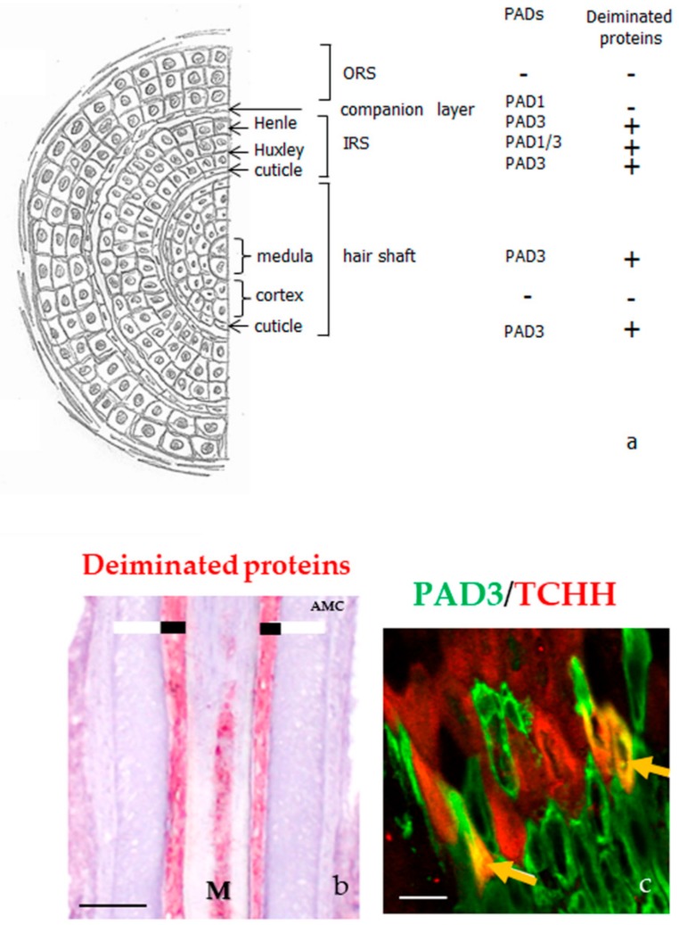
Figure 4.
PADs and deiminated proteins in the hair follicles. (a) Schematic representation of a hair follicle section and detection of both PADs and deiminated proteins, as reported in [26,27]. IRS, inner root sheath; ORS, outer root sheath. (b) Immunodetection of deiminated proteins (red) with the anti-modified citrulline antibody in the medulla (M) and the inner root sheath (large black line) of a normal human hair follicle. The outer root sheath is indicated with a large white line. (c) Immunofluorescence detection of PAD3 (green) and trichohyalin (TCHH, red). A yellow staining (yellow arrows) corresponds to colocation of the two proteins. Bar = 50 µm (b) and 100 µm (c).

