Figure 5.
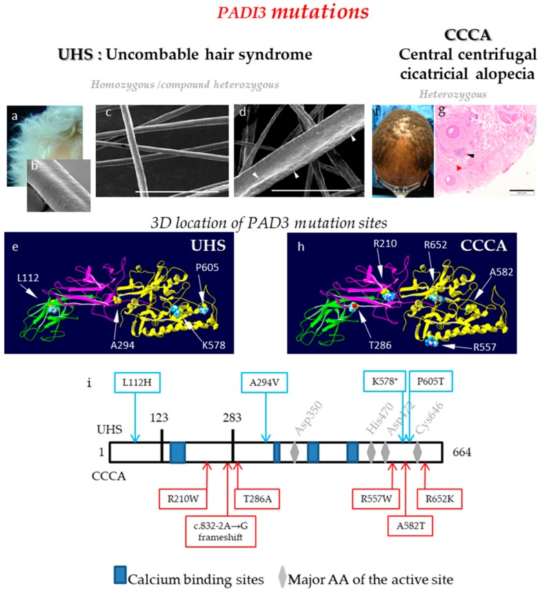
PADs and deimination in hair follicle disorders. (a) Illustration of the macroscopic hair phenotype of an UHS patient. (b-d) Representative scanning electron microscopy observations of the abnormal longitudinal UHS hair shape (b), hair follicles of Padi3 wild type mice; (c) and hair follicles of Padi3 knockout mice (d). Bar = 150 µm. White arrowheads point the abnormalities of the hair shape. (e) Localization of PAD3 amino-acids that are mutated in UHS patients. (f and g) Hair phenotype of a Central Centrifugal Cicatricial Alopecia (CCCA) patient and histological observation. Black and red arrowheads point to inflammatory infiltrate, and fibrosis, respectively. Bar = 200 µm. (h) Localization of PAD3 amino-acids that are mutated in CCCA patients. (i) Position of the pathological mutations on a linear representation of PAD3. Mutations responsible for UHS (top) or CCCA (bottom). The in silico 3D model of PAD3 (e,h) was produced by similarity from the crystallographic data of PAD4 (PDB ID: 1WDA), as previously published [9,16,22].

