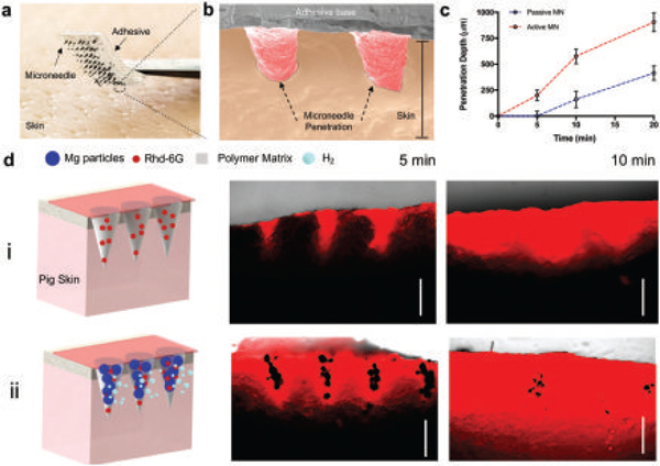
Figure 4.

Evaluation of ex vivo dye release performance of passive and active microneedles. a) Digital photograph of an active microneedle patch (7 × 7 array) loaded with Rh6G before piercing porcine skin. b) Colored SEM image of 2 active microneedles piercing porcine skin. Scale bar, 1 mm. c) Corresponding penetration depth of Rh6G by microneedles at different time points, n = 3. d) Schematic illustrating the experimental set up of both passive and active microneedles penetrating into porcine skin. Passive (i) and active (ii) microneedle arrays are shown, along with fluorescence microscopy cross-section images at different times. Scale bars, 500 μm.
