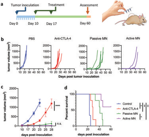
Figure 5.

In vivo melanoma tumor eradication by active microneedles. a) In vivo skin-cancer treatment using anti-CTLA-4 antibodies delivered by active versus passive microneedles and intratumoral injection in the B16F10 dermal melanoma model. b) Tumor volumes growth curve of individual mice and averaged tumor volumes of mice receiving PBS (blue), free anti-CTLA-4 (red), anti-CTLA-4 passive microneedle (MN, green), and anti-CLTA-4 active MN (purple). Data are means ± SEM (n = 3–5). c) Tumor growth over time was compared by two-way ANOVA with Tukey’s test: ****p < 0.0001, n.s. no significant difference. d) Survival rates. Statistical significance was calculated using Log-rank (Mantel–Cox) test: *p < 0.05, **p < 0.01.
