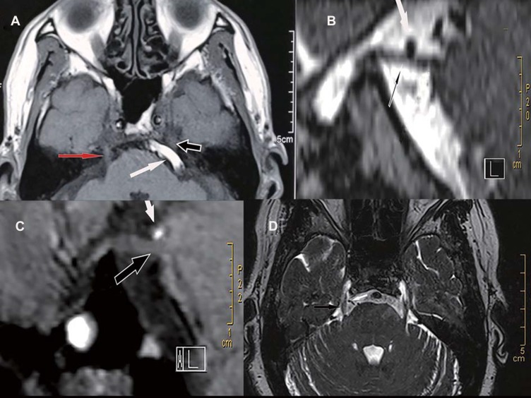
Figure 1.
Preoperative magnetic resonance imaging (MRI) images under different neurovascular compression. (A) The left basilar artery (BA; white arrow) obviously compresses the trigeminal nerve (black arrow) and causes the nerve root to displacement. Note that the trigeminal nerve on the right (red arrow) has no vascular compression and no displacement. (B) The left superior cerebellar artery (SCA; white arrow) obviously compresses the trigeminal nerve (black arrow). (C) The SCA (white arrow) only contacts the trigeminal nerve (black arrow). Note that there is no cerebrospinal fluid visualized between the nerve and the artery. No deformity of the nerve is observed. (D) Only the vein (white arrow) compresses the right trigeminal nerve (black arrow).

