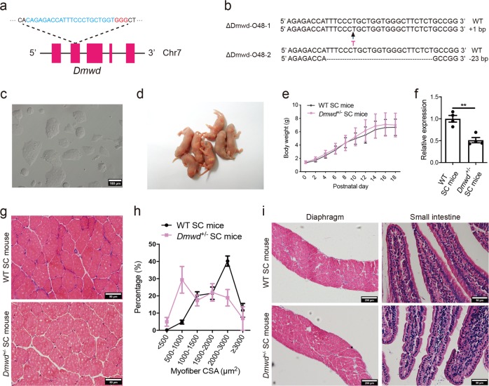
Fig. 2.
Generation of Dmwd+/− SC mice through ICAHCI of haploid cells carrying mutant Dmwd. a Schematic of the sgRNA targeting Dmwd. b Sequences of the Dmwd gene in two cell lines (ΔDmwd-O48-1 and ΔDmwd-O48-2) carrying CRISPR-Cas9-induced gene modifications. c Phase-contrast image of ΔDmwd-O48-1 cell line. Scale bar, 100 μm. d Newborn SC pups generated from ΔDmwd-O48-1 cells. e Average body weight of Dmwd+/− SC mice and WT SC mice (n > 8 per group, means ± SD). f Transcription analysis of Dmwd in TA muscles showed that the transcription level of Dmwd was significantly lower in Dmwd+/− SC mice compared with WT SC mice (n = 4 per group, 4-6-month old). Unpaired Student’s t-test, **P < 0.01. g Representative images of H&E staining of TA muscles from DMWD+/− SC mice and WT SC mice (4-6-month old). Scale bars, 50 μm. h CSA analysis of TA muscle showed muscle wasting in DMWD+/− SC mice (n = 3 per group, 4-6-month old). i Representative images of H&E staining showed normal histological structure of diaphragm and small intestine in DMWD+/− SC mice (4-6-month old). Scale bars, 200 µm for diaphragm; 50 µm for small intestine

