Fig. 3.
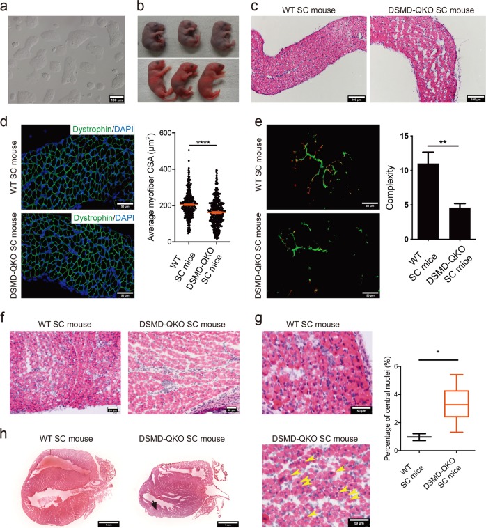
SC mice carrying heterozygous mutations in Dmpk, Six5, Mbnl1 and Dmwd show CDM phenotypes. a Phase-contrast image of cultured ΔDSMD-O48-1 haploid cells. Scale bar, 100 μm. b 23% of newborn DSMD-QKO SC pups died during perinatal period (upper), while others survived (lower). c H&E staining of diaphragm sections from DSMD-QKO and WT SC pups (P1). Scale bars, 100 μm. d Diaphragm CSA analysis of DSMD-QKO and WT SC mice (P1) (n = 3 per group). Unpaired Student’s t-test, ****P < 0.0001. Scale bars, 50 μm. e Immunofluorescent staining of bungarotoxin (red) and neurofilament H (green), and complexity analysis of NMJs of DSMD-QKO and WT SC mice (P1) (WT SC pups, n = 2; DSMD-QKO SC pups, n = 5). Unpaired Student’s t-test, **P < 0.01. Scale bars, 50 μm. f H&E staining of TA muscle sections (P1) showing less myofiber in DSMD-QKO SC mice (2/5). Scale bars, 50 μm. g H&E staining of TA muscle of DSMD-QKO and WT SC (P0) mice and the percentage of myofibers containing centrally located nuclei in TA muscle (n = 3 per group). Yellow arrows indicate the myofibers with central nuclei. Unpaired Student’s t-test, *P < 0.05. Scale bars, 50 μm. h H&E staining of heart cryo-sections (P17) showing left ventricular posterior wall attenuation in DSMD-QKO SC mice (2/7, black arrow). Scale bars, 1 mm

