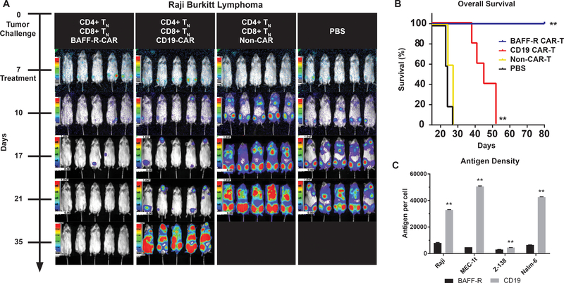
Figure 3. Superiority of BAFF-R versus CD19 CAR T cells in a Burkitt lymphoma model is not due to greater tumor antigen density.
(a) Bioluminescence images of groups of 5 NSG mice following IV tumor challenge (0.5 × 10⁶ cells/mouse) on day 0 with luciferase-expressing Raji cells. 2.5 × 10⁶ activated CD4 TN CAR-T + 106 CD8 TN BAFF-R- or CD19-CAR T cells were infused IV on day 7 as a single dose. Control mice received non-transduced CD4/CD8 T cells from the same donor as an allogeneic control, or PBS. Data are representative of two independent experiments using different donor T cells. (b) Kaplan-Meier plot of overall survival at 80 days is shown. Log-rank test: **P<0.01 compared with all other groups. (c) Calculated cell surface antigen density of BAFF-R and CD19 on lymphoma and leukemia lines stained by PE-conjugated antibodies at saturation. PE per cell (assuming 1 PE per antibody) was calculated against mean fluorescence intensity (MFI) standard curve with BD Quantibrite beads. Data are represented as mean ± s.d. of triplicates. Student’s t-test: **P<0.001 BAFF-R vs. CD19 in corresponding cell line.

