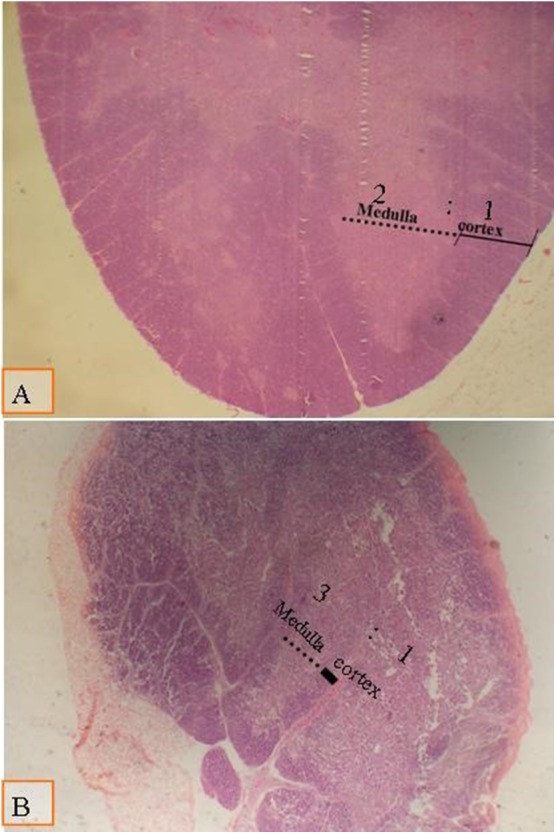
Figure 3.

Micrograph of the thymus stained with H&E (×40) to compare the thymic cortical/medullary ratio: (A) the cortic/medullary ratio is 2:1 as shown in all groups at day 28 of age; (B) the cortic/medullary ration is 3:1 as shown in all groups at day 38 of age, which associated with physiological aged thymic atrophy.
